Cornu Ammonis Region 3 on:
[Wikipedia]
[Google]
[Amazon]

 Hippocampus anatomy describes the physical aspects and properties of the
  Topologically, the surface of a cerebral hemisphere can be regarded as a sphere with an indentation where it attaches to the midbrain. The structures that line the edge of the hole collectively make up the so-called
 Starting at the dentate gyrus and working inward along the S-curve of the hippocampus means traversing a
series of narrow zones. The first of these, the
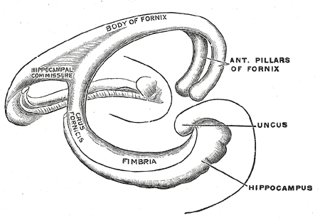
 Fimbria-fornix fibers are the hippocampal and subicular gateway ''to'' and ''from'' subcortical brain regions. Different parts of this system are given different names:
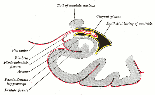
* White myelinated fibers that cover the ''ventricular (deep)'' parts of hippocampus make alveus.
* Fibers that cover the ''temporal'' parts of hippocampus make a fiber bundle that is called fimbria. Going from temporal to septal (dorsal) parts of hippocampus fimbria collects more and more hippocampal and subicular outputs and becomes thicker.
* In the ''midline'' and under the
O-LMR cells
It is also known that extracellular stimulation of ''fimbria'' stimulates CA3 Pyramidal cells antidromically and orthodromically, but it has no impact on dentate granule cells. Each CA1 Pyramidal cell also sends an axonal branch to fimbria.
DG hilar perforant path-associated (HIPP)
an
CA3 trilaminar
cells antidromically.


Schematic Diagram of a Hippocampal Brain Slice
* *
Hippocampus anatomy and connectivity
{{Authority control Hippocampus (brain)

 Hippocampus anatomy describes the physical aspects and properties of the
 hippocampus
The hippocampus (via Latin from Greek , 'seahorse') is a major component of the brain of humans and other vertebrates. Humans and other mammals have two hippocampi, one in each side of the brain. The hippocampus is part of the limbic system, a ...
, a neural structure in the medial temporal lobe
The temporal lobe is one of the four Lobes of the brain, major lobes of the cerebral cortex in the brain of mammals. The temporal lobe is located beneath the lateral fissure on both cerebral hemispheres of the mammalian brain.
The temporal lobe ...
of the brain
A brain is an organ that serves as the center of the nervous system in all vertebrate and most invertebrate animals. It is located in the head, usually close to the sensory organs for senses such as vision. It is the most complex organ in a v ...
. It has a distinctive, curved shape that has been likened to the sea-horse monster of Greek mythology
A major branch of classical mythology, Greek mythology is the body of myths originally told by the Ancient Greece, ancient Greeks, and a genre of Ancient Greek folklore. These stories concern the Cosmogony, origin and Cosmology#Metaphysical co ...
and the ram's horns of Amun
Amun (; also ''Amon'', ''Ammon'', ''Amen''; egy, jmn, reconstructed as (Old Egyptian and early Middle Egyptian) → (later Middle Egyptian) → (Late Egyptian), cop, Ⲁⲙⲟⲩⲛ, Amoun) romanized: ʾmn) was a major ancient Egyptian ...
in Egyptian mythology
Egyptian mythology is the collection of myths from ancient Egypt, which describe the actions of the Egyptian gods as a means of understanding the world around them. The beliefs that these myths express are an important part of ancient Egyptia ...
. This general layout holds across the full range of mammal
Mammals () are a group of vertebrate animals constituting the class Mammalia (), characterized by the presence of mammary glands which in females produce milk for feeding (nursing) their young, a neocortex (a region of the brain), fur or ...
ian species, from hedgehog to human, although the details vary. For example, in the rat, the two hippocampi look similar to a pair of bananas, joined at the stems. In primate
Primates are a diverse order of mammals. They are divided into the strepsirrhines, which include the lemurs, galagos, and lorisids, and the haplorhines, which include the tarsiers and the simians (monkeys and apes, the latter including huma ...
brains, including humans, the portion of the hippocampus near the base of the temporal lobe is much broader than the part at the top. Due to the three-dimensional curvature of this structure, two-dimensional sections such as shown are commonly seen. Neuroimaging
Neuroimaging is the use of quantitative (computational) techniques to study the structure and function of the central nervous system, developed as an objective way of scientifically studying the healthy human brain in a non-invasive manner. Incre ...
pictures can show a number of different shapes, depending on the angle and location of the cut.
 Topologically, the surface of a cerebral hemisphere can be regarded as a sphere with an indentation where it attaches to the midbrain. The structures that line the edge of the hole collectively make up the so-called
 limbic system
The limbic system, also known as the paleomammalian cortex, is a set of brain structures located on both sides of the thalamus, immediately beneath the medial temporal lobe of the cerebrum primarily in the forebrain.Schacter, Daniel L. 2012. ''Ps ...
(Latin ''limbus'' =
''border''), with the hippocampus lining the posterior edge of this hole. These limbic structures include the hippocampus, cingulate cortex
The cingulate cortex is a part of the brain situated in the medial aspect of the cerebral cortex. The cingulate cortex includes the entire cingulate gyrus, which lies immediately above the corpus callosum, and the continuation of this in the ci ...
, olfactory cortex
The olfactory system, or sense of smell, is the sensory system used for smelling (olfaction). Olfaction is one of the special senses, that have directly associated specific organs. Most mammals and reptiles have a main olfactory system and an ac ...
, and amygdala
The amygdala (; plural: amygdalae or amygdalas; also '; Latin from Greek, , ', 'almond', 'tonsil') is one of two almond-shaped clusters of nuclei located deep and medially within the temporal lobes of the brain's cerebrum in complex verteb ...
. Paul MacLean once suggested, as part of his triune brain
The triune brain is a model of the evolution of the vertebrate forebrain and behavior, proposed by the American physician and neuroscientist Paul D. MacLean in the 1960s. The triune brain consists of the reptilian complex (basal ganglia), the p ...
theory, that the limbic structures constitute the neural basis of emotion
Emotions are mental states brought on by neurophysiological changes, variously associated with thoughts, feelings, behavioral responses, and a degree of pleasure or displeasure. There is currently no scientific consensus on a definition. ...
. While most neuroscientists no longer believe in the concept of a unified "limbic system", these regions are highly interconnected and do interact with one another.
Basic hippocampal circuit
 Starting at the dentate gyrus and working inward along the S-curve of the hippocampus means traversing a
series of narrow zones. The first of these, the
 dentate gyrus
The dentate gyrus (DG) is part of the hippocampal formation in the temporal lobe of the brain, which also includes the hippocampus and the subiculum. The dentate gyrus is part of the hippocampal trisynaptic circuit and is thought to contribute ...
(DG), is actually a separate
structure, a tightly packed layer of small granule cell
A granule is a large particle or grain. It can refer to:
* Granule (cell biology), any of several submicroscopic structures, some with explicable origins, others noted only as cell type-specific features of unknown function
** Azurophilic granul ...
s wrapped around the end of the hippocampus proper
The hippocampus proper refers to the actual structure of the hippocampus which is made up of three regions or subfields. The subfields CA1, CA2, and CA3 use the initials of cornu Ammonis, an earlier name of the hippocampus.
Structure
There are ...
, forming a pointed wedge in some cross-sections, a semicircle in others. Next
come a series of ''Cornu Ammonis'' areas: first CA4 (which underlies the dentate gyrus), then CA3, then
a very small zone called CA2
Calcium ions (Ca2+) contribute to the physiology and biochemistry of organisms' cells. They play an important role in signal transduction pathways, where they act as a second messenger, in neurotransmitter release from neurons, in contraction of ...
, then CA1. The CA areas are all filled with densely packed Pyramidal cells
Pyramidal cells, or pyramidal neurons, are a type of multipolar neuron found in areas of the brain including the cerebral cortex, the hippocampus, and the amygdala. Pyramidal neurons are the primary excitation units of the mammalian prefrontal cor ...
similar to those found in the neocortex
The neocortex, also called the neopallium, isocortex, or the six-layered cortex, is a set of layers of the mammalian cerebral cortex involved in higher-order brain functions such as sensory perception, cognition, generation of motor commands, sp ...
. After CA1 comes an area called the subiculum
The subiculum (Latin for "support") is the most inferior component of the hippocampal formation. It lies between the entorhinal cortex and the CA1 subfield of the hippocampus proper.
The subicular complex comprises a set of related structures in ...
. After this comes a pair of ill-defined areas called the presubiculum and parasubiculum, then a
transition to the cortex proper (mostly the entorhinal
The entorhinal cortex (EC) is an area of the brain's allocortex, located in the medial temporal lobe, whose functions include being a widespread network hub for memory, navigation, and the perception of time.Integrating time from experience in the ...
area of the cortex). Most anatomists
use the term "hippocampus proper" to refer to the four CA fields, and hippocampal formation
The hippocampal formation is a compound structure in the Temporal lobe#Medial temporal lobe, medial temporal lobe of the brain. It forms a c-shaped bulge on the floor of the temporal horn of the Lateral ventricles, lateral ventricle. There is no ...
to refer to the hippocampus proper plus dentate gyrus and subiculum.
The major signaling pathways
Signal transduction is the process by which a chemical or physical signal is transmitted through a cell as a series of molecular events, most commonly protein phosphorylation catalyzed by protein kinases, which ultimately results in a cellular ...
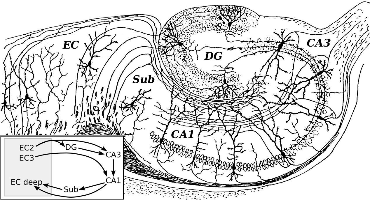
flow through the hippocampus and combine to form a loop. Most external input comes from the adjoining entorhinal cortex
The entorhinal cortex (EC) is an area of the brain's allocortex, located in the medial temporal lobe, whose functions include being a widespread network hub for memory, navigation, and the perception of time.Integrating time from experience in the ...
, via the axons of the so-called perforant path
In the brain, the perforant path or perforant pathway provides a connectional route from the entorhinal cortex to all fields of the hippocampal formation, including the dentate gyrus, all CA fields (including CA1), and the subiculum.
Though it a ...
. These axons arise from layer 2 of the entorhinal cortex (EC), and terminate in the dentate gyrus and CA3. There is also a distinct pathway from layer 3 of the EC directly to CA1, often referred to as the temporoammonic or TA-CA1 pathway. Granule cells of the DG send their axons (called "mossy fibers") to CA3. Pyramidal cells of CA3 send their axons to CA1. Pyramidal cells of CA1 send their axons to the subiculum and deep layers of the EC. Subicular neurons send their axons mainly to the EC. The perforant path-to-dentate gyrus-to-CA3-to-CA1 was called the trisynaptic circuit by Per Andersen, who noted that thin slices could be cut out of the hippocampus perpendicular to its long axis, in a way that preserves all of these connections. This observation was the basis of his ''lamellar hypothesis'', which proposed that the hippocampus can be thought of as a series of parallel strips, operating in a functionally independent way. The lamellar concept is still sometimes considered to be a useful organizing principle, but more recent data, showing extensive longitudinal connections within the hippocampal system, have required it to be substantially modified.
Perforant path input from EC layer II enters the dentate gyrus and is relayed to region CA3 (and to mossy cells, located in the hilus of the dentate gyrus, which then send information to distant portions of the dentate gyrus where the cycle is repeated). Region CA3 combines this input with signals from EC layer II and sends extensive connections within the region and also sends connections to strata radiatum and oriens of ipsilateral and contralateral CA1 regions through a set of fibers called the Schaffer collateral Schaffer collaterals are axon collaterals given off by CA3 pyramidal cells in the hippocampus. These collaterals project to area CA1 of the hippocampus and are an integral part of memory formation and the emotional network of the Papez circuit, and ...
s, and commissural
pathway, respectively. Region CA1 receives input from the CA3 subfield, EC layer III and the nucleus reuniens of the thalamus (which project only to the terminal apical dendritic tufts in the stratum lacunosum-moleculare). In turn, CA1 projects to the subiculum as well as sending information along the aforementioned output paths of the hippocampus. The subiculum is the final stage in the pathway, combining information from the CA1 projection and EC layer III to also send information along the output pathways of the hippocampus.
The hippocampus also receives a number of subcortical inputs. In ''Macaca fascicularis
The crab-eating macaque (''Macaca fascicularis''), also known as the long-tailed macaque and referred to as the cynomolgus monkey in laboratories, is a cercopithecine primate native to Southeast Asia. A species of macaque, the crab-eating macaqu ...
'', these inputs include the amygdala
The amygdala (; plural: amygdalae or amygdalas; also '; Latin from Greek, , ', 'almond', 'tonsil') is one of two almond-shaped clusters of nuclei located deep and medially within the temporal lobes of the brain's cerebrum in complex verteb ...
(specifically the anterior amygdaloid area, the basolateral nucleus, and the periamygdaloid cortex), the medial septum
The medial septal nucleus (MS) is one of the septal nuclei. Neurons in this nucleus give rise to the bulk of efferents from the septal nuclei. A major projection from the medial septal nucleus terminates in the hippocampal formation.
It plays a ro ...
and the diagonal band of Broca
The diagonal band of Broca is one of the basal forebrain structures that are derived from the ventral telencephalon during development. This structure forms the medial margin of the anterior perforated substance. This brain region was described b ...
, the claustrum
The claustrum (Latin, meaning "to close" or "to shut") is a thin, bilateral collection of neurons and supporting glial cells, that connects to cortical (e.g., the pre-frontal cortex) and subcortical regions (e.g., the thalamus) of the brain. It ...
, the substantia innominata
The substantia innominata also innominate substance, or substantia innominata of Meynert (Latin for unnamed substance) is a series of layers in the human brain consisting partly of gray and partly of white matter, which lies below the anterior part ...
and the basal nucleus of Meynert, the thalamus
The thalamus (from Greek θάλαμος, "chamber") is a large mass of gray matter located in the dorsal part of the diencephalon (a division of the forebrain). Nerve fibers project out of the thalamus to the cerebral cortex in all directions, ...
(including the anterior nuclear complex, the laterodorsal nucleus, the paraventricular and parataenial nuclei, the nucleus reuniens, and the nucleus centralis medialis), the lateral preoptic and lateral hypothalamic
The hypothalamus () is a part of the brain that contains a number of small nuclei with a variety of functions. One of the most important functions is to link the nervous system to the endocrine system via the pituitary gland. The hypothalamus i ...
areas, the supramammillary and retromammillary regions, the ventral tegmental area
The ventral tegmental area (VTA) (tegmentum is Latin for ''covering''), also known as the ventral tegmental area of Tsai, or simply ventral tegmentum, is a group of neurons located close to the midline on the floor of the midbrain. The VTA is the ...
, the tegmental reticular fields, the raphe nuclei (the nucleus centralis superior and the dorsal raphe nucleus), the nucleus reticularis tegementi pontis, the periaqueductal gray
The periaqueductal gray (PAG, also known as the central gray) is a brain region that plays a critical role in autonomic function, motivated behavior and behavioural responses to threatening stimuli. PAG is also the primary control center for d ...
, the dorsal tegmental nucleus, and the locus coeruleus
The locus coeruleus () (LC), also spelled locus caeruleus or locus ceruleus, is a nucleus in the pons of the brainstem involved with physiological responses to stress and panic. It is a part of the reticular activating system.
The locus coerule ...
.
The hippocampus also receives direct monosynaptic projections from the cerebellar fastigial nucleus
The fastigial nucleus is located in the cerebellum. It is one of the four deep cerebellar nuclei (the others being the nucleus dentatus, nucleus emboliformis and nucleus globosus), and is grey matter embedded in the white matter of the cerebell ...
.
Major fiber systems in the rat
Angular bundle
These fibers start from the ventral part of entorhinal cortex (EC) and contain commissural (EC◀▶Hippocampus) and Perforant path (excitatory EC▶CA1, and inhibitory EC◀▶CA2) fibers. They travel along the septotemporal axis of the hippocampus. Perforant path fibers, as the name suggests, perforate subiculum before going to the hippocampus (CA fields) and dentate gyrus.Fimbria-fornix pathway
 Fimbria-fornix fibers are the hippocampal and subicular gateway ''to'' and ''from'' subcortical brain regions. Different parts of this system are given different names:
* White myelinated fibers that cover the ''ventricular (deep)'' parts of hippocampus make alveus.
* Fibers that cover the ''temporal'' parts of hippocampus make a fiber bundle that is called fimbria. Going from temporal to septal (dorsal) parts of hippocampus fimbria collects more and more hippocampal and subicular outputs and becomes thicker.
* In the ''midline'' and under the
 corpus callosum
The corpus callosum (Latin for "tough body"), also callosal commissure, is a wide, thick nerve tract, consisting of a flat bundle of commissural fibers, beneath the cerebral cortex in the brain. The corpus callosum is only found in placental mam ...
, these fibers form the fornix.
At the circuit level, the ''alveus'' contains axonal fibers from the DG and from Pyramidal neurons of CA3, CA2, CA1 and subiculum (CA1 ▶ subiculum and CA1 ▶ entorhinal projections) that collect in the temporal hippocampus to form the fimbria/fornix, one of the major outputs of the hippocampus. In the rat, some medial and lateral entorhinal axons (entorhinal ▶ CA1 projection) pass through alveus towards the CA1 stratum lacunosum moleculare without making a significant number of en passant boutons on other CA1 layers (Temporoammonic alvear pathway). Contralateral entorhinal ▶ CA1 projections almost exclusively pass through alveus. The more septal the more ipsilateral entorhinal-CA1 projections that take alvear pathway (instead of perforant path). Although subiculum sends axonal projections to alveus, subiculum ▶ CA1 projection passes through strata oriens and moleculare of subiculum and CA1. Cholinergic and GABAergic projections from MS-DBB to CA1 also pass through Fimbria. Fimbria stimulation leads to cholinergic excitation of CAO-LMR cells
It is also known that extracellular stimulation of ''fimbria'' stimulates CA3 Pyramidal cells antidromically and orthodromically, but it has no impact on dentate granule cells. Each CA1 Pyramidal cell also sends an axonal branch to fimbria.
Hippocampal commissures
Hilar mossy cells and CA3 Pyramidal cells are the main origins of hippocampal commissural fibers. They pass through hippocampal commissures to reach contralateral regions of hippocampus. Hippocampal commissures have dorsal and ventral segments. Dorsal commissural fibers consists mainly ofentorhinal
The entorhinal cortex (EC) is an area of the brain's allocortex, located in the medial temporal lobe, whose functions include being a widespread network hub for memory, navigation, and the perception of time.Integrating time from experience in the ...
and presubicular fibers to or from the hippocampus and dentate gyrus. As a rule of thumb, one could say that each cytoarchitectonic field that contributes to the commissural projection also has a parallel associational fiber that terminates in the ipsilateral hippocampus. The inner molecular layer of dentate gyrus (dendrites of both granule cells and GABAergic interneurons) receives a projection that has both associational and commissural fibers mainly from hilar mossy cells and to some extent from CA3c Pyramidal cells. Because this projection fibers originate from both ipsilateral and contralateral sides of hippocampus they are called associational/commissural projections. In fact, each mossy cell innervates both the ipsilateral and contralateral dentate gyrus. The well known trisynaptic circuit of the hippocampus spans mainly horizontally along the hippocampus. However, associational/commissural fibers, like CA2 Pyramidal cell associational projections, span mainly longitudinally (dorsoventrally) along the hippocampus.
Commissural fibers that originate from CA3 Pyramidal cells go to CA3, CA2 and CA1 regions. Like mossy cells, a single CA3 Pyramidal cell contributes to both commissural and associational fibers, and they terminate on both principal cells and interneurons. A weak commissural projection connects both CA1 regions together. Subiculum has no commissural inputs or outputs. In comparison with rodents, hippocampal commissural connections are much less abundant in the monkey and humans. Although excitatory cells are the main contributors to commissural pathways, a GABAergic component has been reported among their terminals which were traced back to hilus as origin. Stimulation of commissural fibers stimulateDG hilar perforant path-associated (HIPP)
an
CA3 trilaminar
cells antidromically.
Hippocampal cells and layers


Hippocampus proper
Thehippocampus proper
The hippocampus proper refers to the actual structure of the hippocampus which is made up of three regions or subfields. The subfields CA1, CA2, and CA3 use the initials of cornu Ammonis, an earlier name of the hippocampus.
Structure
There are ...
is composed of a number of subfields. Though terminology varies among authors, the terms most frequently used are dentate gyrus
The dentate gyrus (DG) is part of the hippocampal formation in the temporal lobe of the brain, which also includes the hippocampus and the subiculum. The dentate gyrus is part of the hippocampal trisynaptic circuit and is thought to contribute ...
and the cornu ammonis (literally "Ammon
Ammon (Ammonite: 𐤏𐤌𐤍 ''ʻAmān''; he, עַמּוֹן ''ʻAmmōn''; ar, عمّون, ʻAmmūn) was an ancient Semitic-speaking nation occupying the east of the Jordan River, between the torrent valleys of Arnon and Jabbok, in p ...
's horn", abbreviated CA). The dentate gyrus contains the fascia dentata and the hilus, while the CA is differentiated into subfields CA1, CA2, CA3, and CA4.
However, the region known as CA4 is in fact the "deep, polymorphic layer of the dentate gyrus" (as clarified by Theodor Blackstad (1956) and by David Amaral (1978)).
Cut in cross section
Cross section may refer to:
* Cross section (geometry)
** Cross-sectional views in architecture & engineering 3D
*Cross section (geology)
* Cross section (electronics)
* Radar cross section, measure of detectability
* Cross section (physics)
**Abs ...
, the hippocampus is a C-shaped structure that resembles a ram's horns Horns or The Horns may refer to:
* Plural of Horn (instrument), a group of musical instruments all with a horn-shaped bells
* The Horns (Colorado), a summit on Cheyenne Mountain
* ''Horns'' (novel), a dark fantasy novel written in 2010 by Joe Hill ...
. The name ''cornu ammonis'' refers to the Egypt
Egypt ( ar, مصر , ), officially the Arab Republic of Egypt, is a transcontinental country spanning the northeast corner of Africa and southwest corner of Asia via a land bridge formed by the Sinai Peninsula. It is bordered by the Mediter ...
ian deity Amun
Amun (; also ''Amon'', ''Ammon'', ''Amen''; egy, jmn, reconstructed as (Old Egyptian and early Middle Egyptian) → (later Middle Egyptian) → (Late Egyptian), cop, Ⲁⲙⲟⲩⲛ, Amoun) romanized: ʾmn) was a major ancient Egyptian ...
, who has the head of a ram. The horned appearance of the hippocampus is caused by cell density differentials and varying degrees of neuronal fibers.
In rodents, the hippocampus is positioned so that, roughly, one end is near the top of the head (the dorsal or septal end) and one end near the bottom of the head (the ventral or temporal end). As shown in the figure, the structure itself is curved and subfields or regions are defined along the curve, from CA4 through CA1 (only CA3 and CA1 are labeled). The CA regions are also structured depthwise in clearly defined strata (or layers):
* Stratum oriens (str. oriens) is the next layer superficial to the alveus. The cell bodies of inhibitory basket cells and horizontal trilaminar cells, named for their axons innervating three layers—the oriens, Pyramidal, and radiatum are located in this stratum. The basal dendrites
Dendrites (from Greek δένδρον ''déndron'', "tree"), also dendrons, are branched protoplasmic extensions of a nerve cell that propagate the electrochemical stimulation received from other neural cells to the cell body, or soma, of the n ...
of Pyramidal neurons are also found here, where they receive input from other Pyramidal cells, septal
In biology, a septum (Latin for ''something that encloses''; plural septa) is a wall, dividing a cavity or structure into smaller ones. A cavity or structure divided in this way may be referred to as septate.
Examples
Human anatomy
* Interatr ...
fibers and commissural fibers from the contralateral hippocampus (usually recurrent connections, especially in CA3 and CA2.) In rodents the two hippocampi are highly connected, but in primates this commissural connection is much sparser.
* Stratum pyramidale (str. pyr.) contains the cell bodies of the Pyramidal neurons, which are the principal excitatory neurons of the hippocampus. This stratum tends to be one of the more visible strata to the naked eye. In region CA3, this stratum contains synapses from the mossy fibers that course through stratum lucidum. This stratum also contains the cell bodies of many interneurons
Interneurons (also called internuncial neurons, relay neurons, association neurons, connector neurons, intermediate neurons or local circuit neurons) are neurons that connect two brain regions, i.e. not direct motor neurons or sensory neurons. In ...
, including axo-axonic cells, bistratified cell Bistratified ganglion cell can refer to either of two kinds of retinal ganglion cells whose cell body is located in the ganglion cell layer of the retina, the small-field bistratified ganglion cell, also known as small bistratified cell (SBC), and t ...
s, and radial trilaminar cells.
* Stratum lucidum (str. luc.) is one of the thinnest strata in the hippocampus and only found in the CA3 region. Mossy fibers from the dentate gyrus granule cells
A granule is a large particle or grain. It can refer to:
* Granule (cell biology), any of several submicroscopic structures, some with explicable origins, others noted only as cell type-specific features of unknown function
** Azurophilic granul ...
course through this stratum in CA3, though synapses from these fibers can be found in str. pyr.
* Stratum radiatum (str. rad.), like str. oriens, contains septal and commissural fibers. It also contains Schaffer collateral Schaffer collaterals are axon collaterals given off by CA3 pyramidal cells in the hippocampus. These collaterals project to area CA1 of the hippocampus and are an integral part of memory formation and the emotional network of the Papez circuit, and ...
fibers, which are the projection forward from CA3 to CA1. Some interneurons that can be found in more superficial layers can also be found here, including basket cells, bistratified cells, and radial trilaminar cells.
* Stratum lacunosum (str. lac.) is a thin stratum that too contains Schaffer collateral fibers, but it also contains perforant path
In the brain, the perforant path or perforant pathway provides a connectional route from the entorhinal cortex to all fields of the hippocampal formation, including the dentate gyrus, all CA fields (including CA1), and the subiculum.
Though it a ...
fibers from the superficial layers of entorhinal cortex. Due to its small size, it is often grouped together with stratum moleculare into a single stratum called stratum lacunosum-moleculare (str. l-m.).
* Stratum moleculare (str. mol.) is the most superficial stratum in the hippocampus. Here the perforant path fibers form synapses onto the distal, apical dendrites of Pyramidal cells.
* Hippocampal sulcus
The hippocampal sulcus, also known as the hippocampal fissure, is a sulcus that separates the dentate gyrus from the subiculum and the CA1 field in the hippocampus.
Structure
Development
During human fetal development, the hippocampal sulcus ...
(sulc.) or fissure is a cell-free region that separates the CA1 field from the dentate gyrus. Because the phase of recorded theta rhythm
Theta waves generate the theta rhythm, a neural oscillation in the brain that underlies various aspects of cognition and behavior, including learning, memory, and spatial navigation in many animals. It can be recorded using various electrophysi ...
varies systematically through the strata, the sulcus is often used as a fixed reference point for recording EEG
Electroencephalography (EEG) is a method to record an electrogram of the spontaneous electrical activity of the brain. The biosignals detected by EEG have been shown to represent the postsynaptic potentials of pyramidal neurons in the neocortex ...
as it is easily identifiable.
Dentate gyrus
Thedentate gyrus
The dentate gyrus (DG) is part of the hippocampal formation in the temporal lobe of the brain, which also includes the hippocampus and the subiculum. The dentate gyrus is part of the hippocampal trisynaptic circuit and is thought to contribute ...
is composed of a similar series of strata:
* The polymorphic layer (poly. lay.) is the most superficial layer of the dentate gyrus and is often considered a separate subfield (as the hilus). This layer contains many interneurons
Interneurons (also called internuncial neurons, relay neurons, association neurons, connector neurons, intermediate neurons or local circuit neurons) are neurons that connect two brain regions, i.e. not direct motor neurons or sensory neurons. In ...
, and the axons of the dentate granule cells pass through this stratum on the way to CA3.
* Stratum granulosum (str. gr.) contains the cell bodies of the dentate granule cells.
* Stratum moleculare, inner third (str. mol. 1/3) is where both commissural fibers from the contralateral dentate gyrus run and form synapses as well as where inputs from the medial septum
The medial septal nucleus (MS) is one of the septal nuclei. Neurons in this nucleus give rise to the bulk of efferents from the septal nuclei. A major projection from the medial septal nucleus terminates in the hippocampal formation.
It plays a ro ...
terminate, both on the proximal dendrites of the granule cells.
* Stratum moleculare, external two thirds (str. mol. 2/3) is the deepest of the strata, sitting just superficial to the hippocampal sulcus across from stratum moleculare in the CA fields. The perforant path fibers run through this strata, making excitatory synapses onto the distal apical dendrites of granule cells.
An up-to-date knowledge base of hippocampal formation neuronal types, their biomarker profile, active and passive electrophysiological parameters, and connectivity is supported at the ''Hippocampome'' website.
References
External links
Schematic Diagram of a Hippocampal Brain Slice
* *
Hippocampus anatomy and connectivity
{{Authority control Hippocampus (brain)